Why Should You Have An Echocardiography Drummoyne?
Who should undergo echocardiography? Echocardiography Drummoyne is a process in which a patient goes through an ultrasound of the heart, this ultrasound produces sound waves, and a person can have a moving image of the heart. This method contains no radiation and provides more detail than X-rays.
As we discussed above, Echocardiography is an ultrasound of the heart, this ultrasound produces sound waves, and a person can have a moving image of the heart. This method contains no radiation and provides more detail than X-rays. Echocardiography is a good diagnostic tool for heart disease, evaluating the heart’s pumping function, i.e. ejection fraction, in patients with infarction.
Echocardiography is also a good screening test for some heart conditions. However, some situations, such as diseases, require an echocardiographic test. Monitoring of patients with the disease should be accompanied by echocardiography. These are situations in which the echo may affect the patient’s clinical management. Evaluation of valve function, e.g. assessment of systolic or diastolic wall Assessment of left ventricular function, systolic diastolic and regional wall movement, e.g. suspected heart failure in a patient without respiration or preoperative assessment.
 Determine the source of brief cardiac embolism in stroke patients. The role of left ventricular hypertrophy is the assessment of congenital heart disease. These abnormalities are small and usually echo. Contact your doctor for details.
What can I expect during echocardiography?
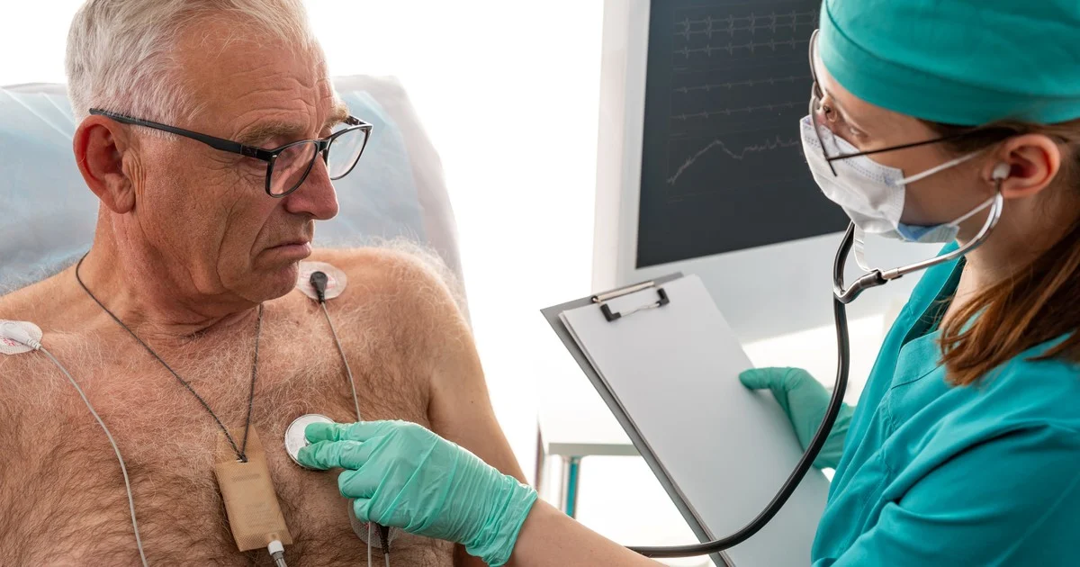
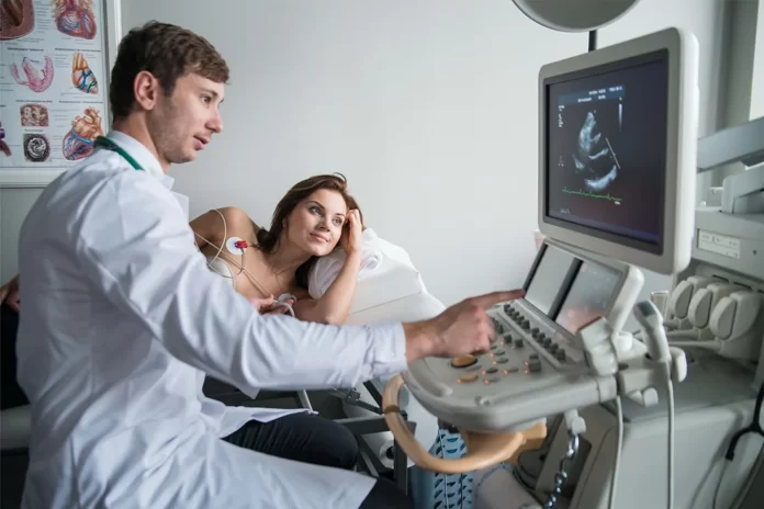
Usually, when you see a cardiologist, your doctor will advise you on echocardiography of your heart. This is an ultrasound of the heart to diagnose various heart diseases. You can use sound waves to move the image of the heart. Echo is a painful and simple procedure without the involvement of any radiation. There are no known risks or risks associated with echocardiography Drummoyne. When you go to the echo room, you are asked to lie on the bed and get up from your waist. Your doctor or echocardiograph will place an electrode in your chest to record an ECG during echocardiography. During this test, a small amount of gel is placed on your breasts, and then a small sensor is placed near your sternum.
The converter produces sound waves to the heart. The echocardiograph can apply some pressure to your chest during the echo. Depending on your technician’s position, you may be asked to turn your side left or right. Your doctor can ask you to hold your breath to take high-quality photos. The echocardiograph then moves the sensor to different parts of the chest to take a picture of the heart from different angles. Sometimes the dye can be injected before it resonates because sometimes the lungs of the body’s ribs prevent sound waves from reaching the heart muscles. If your heart beats too hard, the echo can result badly, so avoid the echo of fast tachycardia. A typical echo develops in approximately 30-45 minutes. If you have lung disease, obesity, dizziness or fatigue, this may result in a longer trial period. During the test, print photographs are taken by an echo machine technician and then examined by a cardiologist.
Drummoyne Advance Cardiology provides the best echocardiography Drummoyne services. We have a team of specialists and experienced cardiologists who are masters in their field. Our main objective is to provide our worthy patients with the best services.
| Other Good Articles to Read |
| Cme Blog Spot |
| Garcias Blogs |
| Yyc Blogs |
| Guiade Blogs |
| Blogs-Hunt |
| Impact-Blog |
| Smarty Blogs |
| Ed Blog |
| Mo Blogs |
| Blogs Em |
| Blog St |


